
Fetal Medicine Services
Motherhood Hospital > Fetal Medicine Services
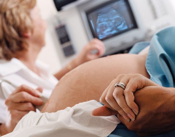
Fetal Ultrasound
Fetal Ultrasound is a sonography done during various phases of pregnancy to see various parts of the intrauterine baby (fetus) like head, heart, spine,…
A. NT Scan
Nuchal Translucency Scan is performed between 11 weeks and 13 weeks of pregnancy. Importance
- To date the pregnancy, we measure the size of the fetus and from this we calculate expected date of delivery.
- To diagnose multiple pregnancy
- To diagnose major fetal anomalies
- To assess the risk of Down’s Syndrome and other chromosomal abnormalities
The NT scan measure the clear fluid behind the neck of the baby. The NT scan is generally offered along with blood tests – double marker test
TEFA Scan Anomaly scan – TIFFA
This is a detailed scan at 18-22 week of pregnancy. During this scan we examine each part of baby including brain, face, spine, heart, stomach, bowel, kidney, bladder, limbs, determine the position of placenta, assess the amount of amniotic fluid, and measures the fetal growth.
If any abnormalities detected significance of finding you can discuss with our consultant fetal medicine at motherhood hospital
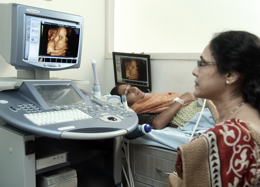
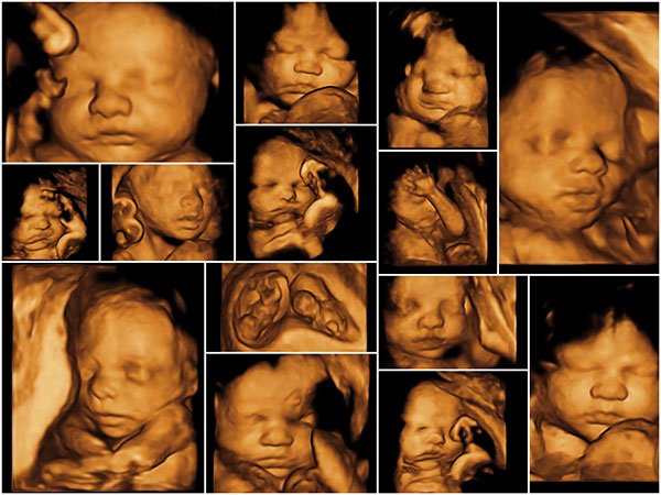
Advanced 3D 4D sonography – 3D/4D sonography
Real-time 3D/4D Sonography in Ahmedabad is possible at Motherhood Hospital. They provide a three-dimensional view of the fetus in motion and are one of the most important modern innovations in the field of ultrasound. The clear view of the baby from all angles allows doctors to detect any congenital abnormalities at an early stage.
The image quality is so clear and sharp that one can get an accurate impression of how a baby’s features look at birth. Motherhood provides live 3D 4D sonography in Sola, Ahmedabad.
Genetic Sonogram
Genetic sonogram is performed when some one is high risk on biochemical screening test. This scan is performed looking for specific ultrasound markers? soft markers which suggestive of possibility of trisomy 21 like venticulomegaly, increased nuchal fold, absent/hypoplastic nasal bone, echogenic cardiac focci, abrrent right subclavian artery(ARSA), echogenic bowel, pyelectasis, short femur.
This soft markers are checked during this scan which either increase or decrease the risk of having trisomy 21 baby. This scan usually performed between 17 to 22 week of gestation along with detailed anatomical evaluation of baby.
Fetal ECHO
Fetal ECHO is a specialised detailed scan of your baby s heart. this scan generally look for heart defect done between 18 to 24 week of pregnancy
Specific scan of fetal heart required in case of patient having history of congenital heart disease, diabetes mellitus, those who are on antiepileptic drug increased NT in NT scan.
Fetal growth and Doppler scan
This scan is usually done between 28 to 36 week of pregnancy. During this scan measurement of baby’ s head, abdomen and thigh bone are taken and estimated baby’s weight is calculated. Also the baby s movement fluid around the baby placental position and blood flow to the baby and palcenta are checked by colour ultrasound.
Multiple Pregnancy scan
Prenatal Screening
They include some blood tests with an ultrasound at 11 week -13 week 6 days. According to the results calculation of having a baby with down’s syndrome and other trisomy and structural abnormality is done. This tests are only screening test they only give chance of your having that chromosomally abnormal baby for confirmation one has to undergo CVS or Amniocentesis.
Aneuploidy Scan/Down’s Syndrome Screening (NT Scan, Biochemical markers, Combined markers, Quadruple, Genetic Sonogram)
Down’s syndrome screening.
Every women has a risk of having baby with down syndrome. This risk either can be decreased or increased by this screening test. After this screening test if any women come under high risk for having down s syndrome she has to undergo diagnostic test for confirmation.
Double marker test / combined 1st trimester screening.
Double marker test include measurement of free B-HCG and PAPA-A in maternal blood . Generally this double marker is used as combine first trimester screening in which along with maternal blood level of PAPAA and free B-HCG, ultrasonography assessment of fetus including nuchal translucency, nasal bone, ductous venosusPI and tricuspid regurgitation done and combined risk of aneuploidy is calculated according to the standard of fetal medicine foundation uk.
Non invasive cell free fetal DNA test(NIPT)
During pregnancy some fetal DNA crosses the placenta and comes to the mother’s blood stream. In this test generally this fetal DNA separated from mothers blood and chromosomal evaluation of baby done . This test is non invasive as its done from mothers blood. This is a screening test with highly sensitive up to 99 %. But one has to undergo diagnostic test for confirmation if high risk comes in report.
Quadruple marker
It includes maternal blood measurements of Inhibin, B HCG, estradiol, and alpha-fetoprotein. This generally combines along with genetic sonogram.
Preeclampsia Screening
- Preeclampsia is a pregnancy induced high blood pressure and is one of the most common life threatening condition during pregnancy.
- This high blood pressure can affect the mothers health and growth of the baby also
- First trimester can identify the women at high risk for for preeclampsia this can improve the pregnancy outcome by appropriate monitoring of fetus and mother and early detection of any affection of baby and timely delivery at appropriate place .
- Ethnicity, first pregnancy ,High BMI ,any previous pregnancy or family history of preeclampsia
- All this factors affect your having risk of preeclampsia.
- In preeclampsia screening include ultrasound measurement of placental blood flow at 11-13 week scan along with history , maternal mean arterial blood pressure with or without blood test.
Preterm Labour risk assessment
- Preterm labour screening
- Preterm labour is when u have started labour pains and open the cervix before 37 completed weeks gestation leads to premature delivery of baby.
- There are many risk factors for preterm labour like poor nutrition , infection, smoking , previous pregnancy having h/o of preterm, cervical surgeries overdistention of uterus, low socioeconomic class etc..
- Because of preterm labour when baby born before 34 week their lungs are still immature may having breathing problems and may require ICU. Also this baby are high risk for infections and feeding problem also.
- Cervical screening by transvaginal sonography done at 11-13+ 6 day and during 19 -22 week anomaly scan. The length of cervix is measured at midsagittal plane to assess the risk of preterm labour. Normally cervix measured more than 25 mm.
Invasive Procedures and fetal therapy
Chorionic Villi Sampling (CVS)
CVS involves the examination of placental tissue (chorionic villi). Both baby and placenta originate from the same cell so the chromosomes of the a placenta are the same as baby.
Amniocentesis
Amniocentesis involves drawing a small amount of amniotic fluid from mother’s womb, surrounding the baby under ultrasound guidance this is generally performed between 17-22 week after detailed ultrasound of the baby. During this procedure exact location of placenta, the quantity of amniotic fluid and timmining of the procedures are determined. Amniotic fluid generally use for chromosomal analysis of fetus, and also in certain viral infections
Cordocentesis
Also known as fetal blood sampling(FBS) and PUBS (percutaneous blood sampling) , a method which includes collection of fetal blood directly from umblical cord under ultrasound gidence. This is used generally in utero complications like , fetal anemia , fetal infections and any other chromosomal defects . it also helps in treating fetus with anemia by in utero blood transfusion. It can help the expectant mothers prepare for a special needs child, and some cases helps parents to decide whether to carry the child decide whether to carry the child to term.
Fetal reduction
Its generally done in multiple pregnancy when there is 3 or more foetuses . the procedure is done at 11 weeks of pregnancy. After cleansing the mothers abdomen with antiseptic solution , a long thin needle is inserted in mothers womb by ultrasound guidance, injection is directly given to the selected fetus which immediately stops its heart activity . procedure is without any bleeding or pain
Fetal Blood Transfusion
This is a highly specialised area of medical practice requiring close collaboration between experts in fetal medicine, Haematology & Blood transfusion, and rapid access to blood counting. The most common indications for intrauterine transfusion (IUT) are red cells for prevention and treatment of fetal anaemia due to haemolytic disease of the fetus & newborn (HDFN) or parvovirus infection and platelets for neonatal alloimmune thrombocytopenia (NAIT).

Genetic Counselling
Genetic counselling provides information and support to people who are or having at risk for genetic disorders. The counselling may be for yourself or for family member, or u may get it when you are planning to have expecting baby.